
Diagnosis
Oncological Risk Assessment: A Strategy for Your Safety
Skin cancer (including melanoma, basal cell, and squamous cell carcinoma) is one of the few oncological diseases that can be detected at the earliest stage without invasive interventions. At VIP Clinic, oncological risk assessment combines the expertise of medical specialists and artificial intelligence technologies.
What does the oncological risk assessment program include?
Collection of oncological history and genetic profile The doctor analyzes your heredity, history of sunburns, Fitzpatrick skin type, and lifestyle characteristics (frequency of flights, visits to countries with high insolation index). This allows us to determine your personal risk group.
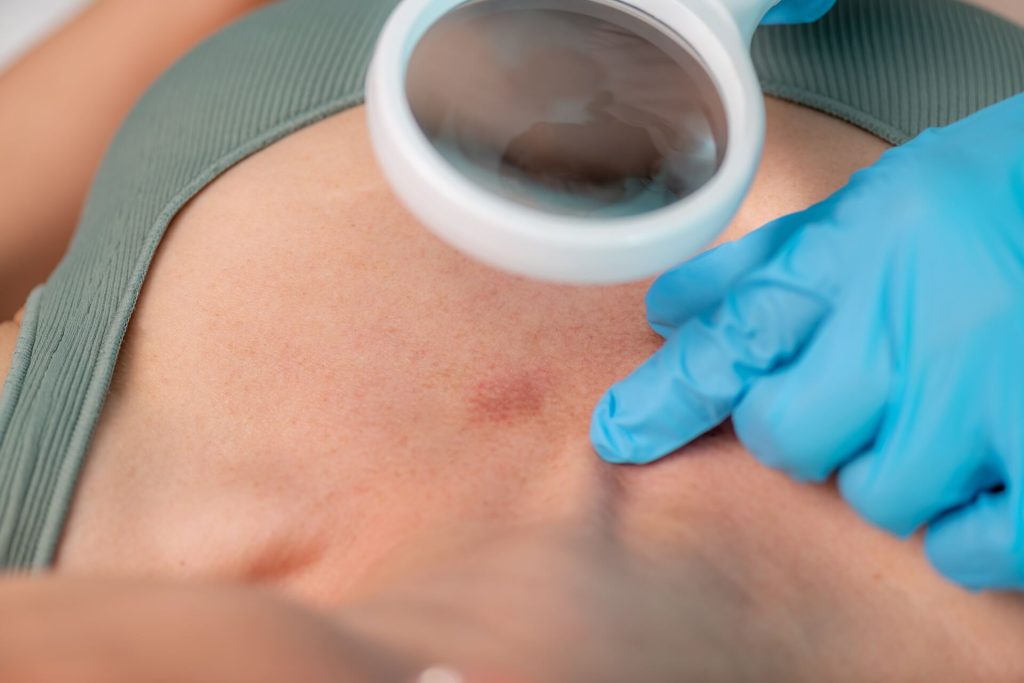
Total Screening (Skin Scan) The doctor examines not only the formations that concern you but also the entire skin surface, including hard-to-reach areas (scalp, interdigital spaces, mucous membranes). We look for the so-called “ugly duckling” sign – a mole that stands out from the rest.
Intelligent Analysis of Formations If suspicious elements are detected, we use the FotoFinder digital system. It analyzes the morphological structure of the neoplasm, evaluating it by dozens of parameters (asymmetry, borders, pigment inclusions, vascular pattern).
Development of a Monitoring Plan Based on the results of the examination, you receive a clear conclusion:
Which formations are absolutely benign.
Which require observation (follow-up photo in 3-6 months).
Which formations require immediate removal with histological analysis.
Who needs annual oncological screening?
We recommend professional risk assessment if at least one of the following applies to you:
Family history: Cases of skin cancer in close relatives.
Phototype: Fair skin, eyes, and hair, presence of freckles.
Quantity: More than 50 moles on the body or atypical nevi (large, irregularly shaped).
Trauma: A mole is located in an area of constant clothing friction or has been damaged.
Changes: You have noticed that a formation has started to itch, bleed, darken, or grow rapidly.
Why is it important to do this specifically at VIP Clinic?
Double control: The conclusion is based on the opinion of an experienced dermato-oncologist and neural network analysis.
Data Archiving: We save a “digital footprint” of each of your formations. This allows us to notice dynamics that cannot be captured during a regular examination.
Histological verification: If a formation requires removal, we perform it within the clinic with mandatory submission of the material to leading pathomorphological laboratories.
Your result: Complete confidence in the safety of every mole and a clear understanding of how to protect your skin in the future.
Patient Memo: The “ABCDE” Rule
We also recommend self-examination, paying attention to 5 signs:
A (Asymmetry) — Asymmetry: one half of the mole does not resemble the other.
B (Border) — Border: irregular, blurred, or notched edges.
C (Color) — Color: presence of several shades (black, brown, gray, red) in one spot.
D (Diameter) — Diameter: formation size greater than 6 mm (the size of a pencil eraser).
E (Evolving) — Evolution: any changes in color, size, or shape over a short period.
Have questions?
Start your journey to health and self-confidence with a consultation with our specialized experts.